This is an old revision of the document!
This page aims to collect together Derriford's suggested pathways for incidental findings commonly seen on general imaging. All the guidance below has been reviewed with local consultants and efforts are made to keep everything up to date, but may be out of date and represents guidance only. It may not fit your patient's specific circumstances and if you have any concerns it is always best to ask a friend.
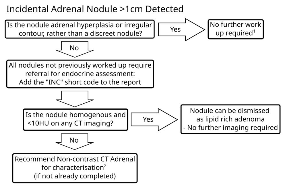
A significant minority of adrenal nodules referred for further assessment are assessed as adrenal hyperplasia or a slightly irregular contour to the adrenal rather than adrenal nodule - is there a clear, discreet nodule distinct from the background adrenal gland?
The most recent guidance does not recommend triple phase adrenal CT in the initial characterisation of adrenal nodules - non-contrast CT is sufficient. Adrenal washout studies are probably best reserved for the adrenal MDT.
This flowchart should only be applied where the detected lesion is truly incidental - in the context of cancer any nodule should be considered a metastasis until proven otherwise.
INC short code text:
This study demonstrates an adrenal incidentaloma > 10 mm in size. If clinically appropriate and not previously investigated, please consider a referral to the adrenal pathway (plh-tr.endocrinesecs@nhs.net).
Underlying guidance:
Last reviewed 14/09/2026
Brain and Spine Tumours
Unless you are confident and happy to dismiss, it is recommended that clinicians are directed to refer all incidentally detected brain and spine tumours to the CNS tumour MDT with the following text inserted into the report:
This imaging has demonstrated a Brain or Spine tumour. If this is a new tumour or recurrence of previous disease then the patient should be referred to the Brain and Spine Tumour MDT Co-ordinator based at Derriford Hospital, Plymouth. To make a referral please email rk9cancerservices@nhs.net and a referral proforma will be sent to you by email for completion. Please arrange an MRI of the brain if clinically appropriate in the interim. If it is a clinical presentation needing urgent attention please contact the on-call Neurosurgery Registrar through 0845 155 8155.
Last reviewed 13/09/2026
Unless you are confident that you can dismiss, it is recommended that all suspicious breast lesions are flagged up to breast radiology, with a comment in the report to inform the clinicians that this has been done. To to get breast radiology input, use the breast radiology email at:
plh-tr.breastradiologyqueries@nhs.net
Breast radiology has access to screening imaging that is not generally available on PACS that may enable them to dismiss the lesion seen, and a significant portion are dismissed on further review, so it is not recommended to refer directly to breast clinic.
Last reviewed 13/09/2026

Underlying guidance:
Last reviewed 13/09/2026

